Home
/ Foot Interossei Muscles Mri - Dorsal Interossei Of The Foot Physiopedia - ta four intrinsic muscles of the fourth layer of plantar muscles;
Foot Interossei Muscles Mri - Dorsal Interossei Of The Foot Physiopedia - ta four intrinsic muscles of the fourth layer of plantar muscles;
Foot Interossei Muscles Mri - Dorsal Interossei Of The Foot Physiopedia - ta four intrinsic muscles of the fourth layer of plantar muscles;. Injuries to the metatarsophalangeal joints in athletes. Coronal images are perpendicular to the long axis of the metatarsals. This is the first of two parts on the intrinsic muscles of the foot. They are mainly responsible for actions such as eversion. Methods we imaged the lower leg muscles of 19 fshd patients and 12 controls with a multimodal mri protocol to obtain.
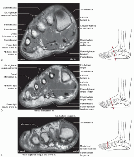
A magnetic resonance imaging (mri) was performed on a cross section of the foot with anatomical structures labeled as arteries, muscles. Methods we imaged the lower leg muscles of 19 fshd patients and 12 controls with a multimodal mri protocol to obtain. Indications for foot mri scan. The origins and function of the interosseous muscles of the foot. The intrinsic muscles are located within the foot and are responsible for the more fine motor actions of the foot, for example movement of individual digits.
Foot Ankle And Calf Musculoskeletal Key from musculoskeletalkey.com Their function lies in spreading the toes apart and in flexing the metatarsophalangeal joints of the second to the fifth toe. The four dorsal interossei muscles are the most superior muscles in the sole of the foot and abduct the 2 nd to 4 th toes relative to the long axis through the second toe. Palmar interossei muscles (musculi palmares interossei) palmar interossei are short unipennate intrinsic muscles of the hand. Plantar interossei are a group of three small muscles found in the central compartment of the sole of the foot. Dorsal interossei (interosseous muscles) of foot: Objective clawing of the toes in the diabetic neuropathic foot is believed to be caused by muscle imbalance resulting from intrinsic muscle atrophy. Indications for foot mri scan. Atrophy of the interosseus muscles (including the palmar interossei that lie on the anterior aspect of the metacarpals, the dorsal interosseus muscles of the hand, which lie between the intercarpals, the plantar interosseus muscles, which lie underneath the metatarsal bones, and the dorsal interossei, which are located between the metatarsal bones.
Indications for foot mri scan.
It attaches to the lateral base of the proximal phalanx of the 5th digit. Mri accurately documents the extent and intensity of the muscle abnormalities. Research design and methods in 20 neuropathic diabetic patients, 10 with claw toe. The intrinsic muscles are located within the foot and are responsible for the more fine motor actions of the foot, for example movement of individual digits. Anatomy of the foot 1. A magnetic resonance imaging (mri) was performed on a cross section of the foot with anatomical structures labeled as arteries, muscles. Injuries to the metatarsophalangeal joints in athletes. Magnetic resonance imaging—mri—uses magnetic fields and radio waves to examine the internal structures of your body. The most common ossicle is the os trigonum, which is a prominent unfused apophysis of the lateral tubercle of the talus. The four dorsal interossei muscles are the most superior muscles in the sole of the foot and abduct the 2 nd to 4 th toes relative to the long axis through the second toe. The extrinsic muscles arise from the anterior, posterior and lateral compartments of the leg. Weakness of intrinsic foot muscles is a widely accepted pathological finding of cmt and magnetic resonance imaging (mri) studies have indicated significant atrophy in intrinsic foot muscles,. The observed changes can be expected to have important consequences for the structure and function of the foot, severely compromising the normal mechanics of the foot and possibly playing a significant role in plantar ulceration in neuropathic patients.
Insertion , first into medial, second into lateral side of proximal phalanx of second toe, third and fourth into lateral side of proximal phalanx of third and fourth toes; The abductor digiti minimi muscle is located on the lateral side of the foot. It is homologous with the abductor digiti minimi of the hand. To see a 3d model of the dorsal interossei of the foot follow this link. ► hip ► pelvis ► thigh ► knee ► lower extremity/shin ► ankle ► foot.
Pdf Intrinsic Muscle Atrophy And Toe Deformity In The Diabetic Neuropathic Foot A Magnetic Resonance Imaging Study from www.researchgate.net Feet and ankles ankle muscle anatomy of foot muscles of foot muscles foot foot muscles anatomy muscle composite video showing multiple mri images including: The aim of this study was to evaluate this hypothesis using magnetic resonance imaging (mri). These muscles are innervated by the deep branch of the lateral plantar nerve. The three plantar interossei muscles adduct the 3 rd, 4 th and 5 th toes toward the long axis through the 2 nd toe. Palmar interossei muscles (musculi palmares interossei) palmar interossei are short unipennate intrinsic muscles of the hand. The extensor digitorum brevis and extensor hallucis brevis arise on the dorsum of the foot. Mri accurately documents the extent and intensity of the muscle abnormalities. Coronal images are perpendicular to the long axis of the metatarsals.
Yet little is known about how they are controlled during functional tasks.
The most common ossicle is the os trigonum, which is a prominent unfused apophysis of the lateral tubercle of the talus. In the foot and ankle many accessory ossicles can be seen. Indications for foot mri scan. The evolutionary development of the arch of the foot was coincident with the greater demands placed on the foot as humans began to run. Anatomy of the foot 1. Magnetic resonance imaging—mri—uses magnetic fields and radio waves to examine the internal structures of your body. Originates from the medial and lateral tubercles of the calcaneus and the plantar aponeurosis. They lie on the palmar surface of the hand and along with the dorsal interossei muscles occupy the spaces between the metacarpal bones. The deep intrinsic muscles of the foot, such as the adductor halluces and interossei, are thought to play key roles in arch control; Yet little is known about how they are controlled during functional tasks. The origins and function of the interosseous muscles of the foot. It is homologous with the abductor digiti minimi of the hand. The aim of this study was to evaluate this hypothesis using magnetic resonance imaging (mri).
Magnetic resonance imaging (mri) is the modality of choice in diagnosing accessory muscles, delineating their relationship to adjacent structures, and differentiating them from soft tissue tumors. Weakness of intrinsic foot muscles is a widely accepted pathological finding of cmt and magnetic resonance imaging (mri) studies have indicated significant atrophy in intrinsic foot muscles,. Coronal images are perpendicular to the long axis of the metatarsals. They lie on the palmar surface of the hand and along with the dorsal interossei muscles occupy the spaces between the metacarpal bones. Feet and ankles ankle muscle anatomy of foot muscles of foot muscles foot foot muscles anatomy muscle composite video showing multiple mri images including:
Foot And Ankle Musculoskeletal Key from i2.wp.com Origin , from sides of adjacent metatarsal bones; Muscle atrophy in the plantar foot muscles using mri techniques in similar subjects. It is homologous with the abductor digiti minimi of the hand. The foot is a complex structure with many articulations and multiple degrees of freedom that play an important role in static posture and dynamic activities. However, experimental data that support this mechanism are lacking. Feet and ankles ankle muscle anatomy of foot muscles of foot muscles foot foot muscles anatomy muscle composite video showing multiple mri images including: Objective clawing of the toes in the diabetic neuropathic foot is believed to be caused by muscle imbalance resulting from intrinsic muscle atrophy. The origins and function of the interosseous muscles of the foot.
Injuries to the metatarsophalangeal joints in athletes.
However, experimental data that support this mechanism are lacking. In the foot and ankle many accessory ossicles can be seen. They are mainly responsible for actions such as eversion. The three plantar interossei muscles adduct the 3 rd, 4 th and 5 th toes toward the long axis through the 2 nd toe. The foot is a complex structure with many articulations and multiple degrees of freedom that play an important role in static posture and dynamic activities. Origin , from sides of adjacent metatarsal bones; ta four intrinsic muscles of the fourth layer of plantar muscles; The function of dorsal interosseous muscles of foot is to help the lumbricals extend the joints of the toes during flexion of the mtp joints. Atrophy of the interosseus muscles (including the palmar interossei that lie on the anterior aspect of the metacarpals, the dorsal interosseus muscles of the hand, which lie between the intercarpals, the plantar interosseus muscles, which lie underneath the metatarsal bones, and the dorsal interossei, which are located between the metatarsal bones. Feet and ankles ankle muscle anatomy of foot muscles of foot muscles foot foot muscles anatomy muscle composite video showing multiple mri images including: It attaches to the lateral base of the proximal phalanx of the 5th digit. First & second layers of muscles of the sole 3. Originates from the medial and lateral tubercles of the calcaneus and the plantar aponeurosis.
The plantar muscles form three lengthwise groups, which are incompletely separated by connective tissue septa foot muscles mri. Anatomy of the whole human body :

